Ultrasound
Ultrasound at ZP
Ultrasounds, or sonograms, are performed at every ZP location. Our state-of-the-art high resolution ultrasound systems obtain images of internal organs and other soft-tissue structures inside the body. Our certified medical sonographers are dedicated to taking the time necessary to provide quality service to all of our patients. Ultrasound is a safe and painless test that uses high frequency sound waves to produce images. The sound waves cannot be heard or felt. Still and moving real-time images can be captured during an ultrasound.

Learn About Different Types of Ultrasound Exams
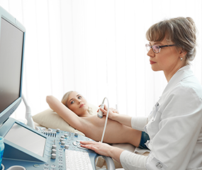 Breast ultrasound (or breast sonogram) is one of the safest and most decisive imaging options available today. They are useful for evaluating nodules, lumps, dense breasts, and more and can differentiate between fluid-filled cysts and solid masses. Commonly they are used along with mammograms to screen for breast cancer or as a guide for biopsies.
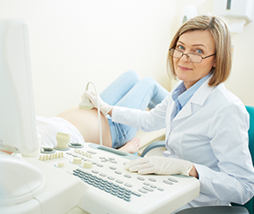
Benefits of Ultrasound
There are no risks because it is non-invasive and doesn’t use radiation
Ultrasound is widely available and less expensive than other imaging methods
Gives clear pictures of soft tissues that don’t show up well on x-ray images
Provides real-time imaging, making it a great tool for guiding minimally-invasive procedures
Tophone® at ZP
Every ZP location uses trophon® on ultrasound probes. Trophon® is a breakthrough disinfection technology for ultrasound probe reprocessing. With the ever increasing challenges in the fight against the spread of Healthcare Acquired Infections (HAIs), trophon’s powerful disinfection technology is setting a new benchmark in protecting patients. At the same time it safeguards staff and the environment from the dangerous hazards and toxic side effects of traditional disinfection methods.
How it Works
Ultrasound imaging uses the same principles of SONAR that were developed during World War I for tracking submarines. It was not used for medical purposes until the late 1940’s.
To produce images, a transducer, or probe, is placed on your skin and pulses of sound waves are sent through your body. As the sound waves pass through the body, they produce echoes which are received by the transducer and sent to the computer. The echoes are analyzed and converted into images, which in turn create real-time pictures on the monitor. This helps to determine the shape, size, and composition of organs and tissues.
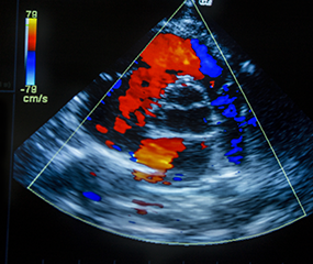
Since ultrasound records images in real time, it is especially useful for examining blood flow and guiding needle biopsy procedures.
During the Test
Depending on the area being studied, you may be asked to change into a gown.
Before the exam starts, the ultrasound technologist will confirm that any special preparation necessary was followed. You will then be asked to lie down on a comfortably padded examination table.
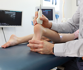
A small amount of gel will be placed on the area being examined. This gel is harmless and can be easily wiped clean after the exam. The gel prevents any air from getting between the transducer (ultrasound probe) and your skin. This direct contact between the probe and your skin helps the transducer deliver sound waves into your body efficiently.
The ultrasound technologist will place the transducer gently on your skin where the gel was applied and move the probe around slowly. Changing the direction or the angle of the probe allows the sonographer to get the best possible images of the organ or tissue being examined.
Preparations
Aortic/Abdominal
Nothing to eat, drink, chew, or smoke for six hours prior to your exam.
Breast/Scrotal/Thyroid
No preparation required.
Color Flow Doppler
No preparation required.
Pelvic/Obstetrical
A full bladder is necessary for the exam. Have breakfast and/or lunch. Women must drink at least 32 oz. of water, finishing one hour prior to your exam. Men must drink at least 16 oz. of water, finishing one hour prior to your exam. Do not empty your bladder.
Prostate
Take a fleet enema at least one hour prior to the exam. Have nothing to eat or drink after the fleet enema.
Renal
Drink a 16 oz. glass of water one hour prior to study. Do not void.
Renal Arterial Study
Have nothing to eat, drink, chew, or smoke for six hours prior to your exam. In addition, consult your physician before taking Gas-X one hour before the exam.
Why Choose Zwanger Pesiri?
Zwanger-Pesiri Radiology brings world-class expertise to the Long Island community. Our subspecialty-trained radiologists are Board Certified by the American Board of Radiology with fellowship training in a variety of specialties. They are highly-skilled, highly-knowledgeable, and make patient care a priority. To learn more, contact us today.